Compound screening in cell-based models of tau inclusion formation: Comparison of primary neuron and HEK293 cell assays
Crowe A, Henderson MJ, Anderson J, Titus SA, Zakharov A, Simeonov A, Buist A, Delay C, Moechars D, Trojanowski JQ, Lee VM, Brunden KR
Journal of Biological Chemistry - vol. 295 4001-4013 (2020)
Journal of Biological Chemistry
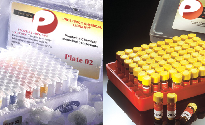
The hallmark pathological features of Alzheimer’s disease (AD) brains are senile plaques, comprising β-amyloid (Aβ) peptides, and neuronal inclusions formed from tau protein. These plaques form 10 -20 years before AD symptom onset, whereas robust tau pathology is more closely associated with symptoms and correlates with cognitive status. This temporal sequence of AD pathology development, coupled with repeated clinical failures of Aβ-directed drugs, suggests that molecules that reduce tau inclusions have therapeutic potential. Few tau-directed drugs are presently in clinical testing, in part because of the difficulty in identifying molecules that reduce tau inclusions. We describe here two cell-based assays of tau inclusion formation that we employed to screen for compounds that inhibit tau pathology: a HEK293 cell-based tau overexpression assay, and a primary rat cortical neuron assay with physiological tau expression. Screening a collection of ~3500 pharmaceutical compounds with the HEK293 cell tau aggregation assay, we obtained only a low number of hit compounds. Moreover, these compounds generally failed to inhibit tau inclusion formation in the cortical neuron assay. We then screened the Prestwick library of mostly approved drugs in the cortical neuron assay, leading to the identification of a greater number of tau inclusion inhibitors. These included four dopamine D2 receptor antagonists, with D2 receptors having previously been suggested to regulate tau inclusions in a Caenorhabditis elegans model. These results suggest that neurons, the cells most affected by tau pathology in AD, are very suitable for screening for tau inclusion inhibitors.


