
High-Throughput Image-Based Aggresome Quantification
Lesire L, Chaput L, Cruz De Casas P, Rousseau F, Piveteau C, Dumont J, Pointu D, Déprez B, Leroux F
SLAS Discovery 1-9 (2020)
SLAS Discovery
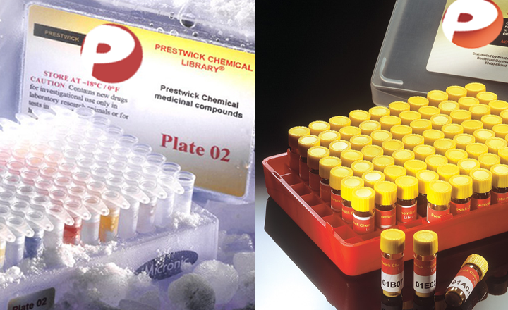
Aggresomes are subcellular perinuclear structures where misfolded proteins accumulate by retrograde transport on microtubules. Different methods are available to monitor aggresome formation, but they are often laborious, time-consuming, and not quantitative. Proteostat is a red fluorescent molecular rotor dye, which becomes brightly fluorescent when it binds to protein aggregates. As this reagent was previously validated to detect aggresomes, we have miniaturized its use in 384-well plates and developed a method for high-throughput imaging and quantification of aggresomes. Two different image analysis methods, including one with machine learning, were evaluated. They lead to similar robust data to quantify cells having aggresome, with satisfactory Z′ factor values and reproducible EC50 values for compounds known to induce aggresome formation, like proteasome inhibitors. We demonstrated the relevance of this phenotypic assay by screening a chemical library of 1280 compounds to find aggresome modulators. We obtained hits that present similarities in their structural and physicochemical properties. Interestingly, some of them were previously described to modulate autophagy, which could explain their effect on aggresome structures. In summary, we have optimized and validated the Proteostat detection reagent to easily measure aggresome formation in a miniaturized, automated, quantitative, and high-content assay. This assay can be used at low, middle, or high throughput to quantify changes in aggresome formation that could help in the understanding of chemical compound activity in pathologies such as protein misfolding disorders or cancer.